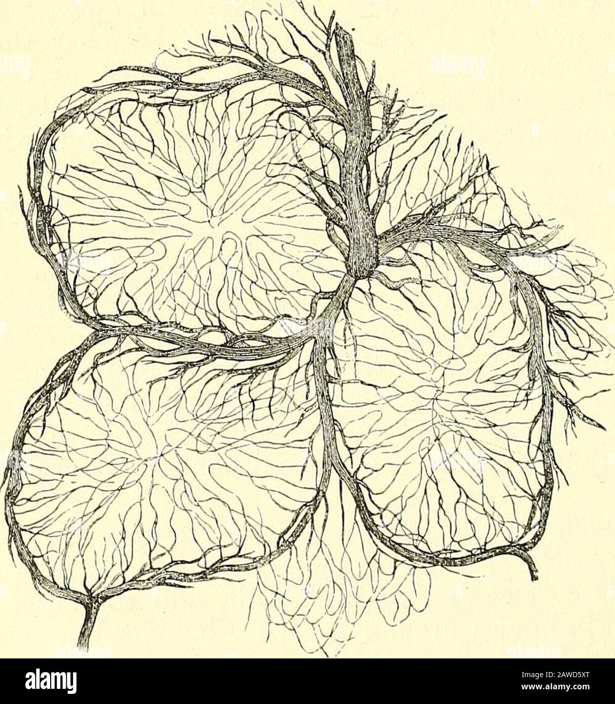
Les éléments de l'anatomie de Quain . emains étant souvent indi- ™ -•—...-:^ . -^ .—-^ catés par coloration foncée de la muqueuse. Vaisseaux et Nerfs.—les branches de l'artère mésentérique, ayant reachedla bordure attachée de l'intestin, passent autour de ses côtés, se divisant en de nombreuses figues. 530. Fig. 530.—Partie d'un patch DE Peyers INJECTÉ(de Kolliker). MAG- KIFIED. Le dessin a été takenfrom une préparation faite byFrey de l'intestin de therabbit. Il représente les vaisseaux sanguins siu-roimdingBlood dans l'introor de trois lymphoidnodosités. Ramifications an
1528 x 1635 px | 25,9 x 27,7 cm | 10,2 x 10,9 inches | 150dpi
Informations supplémentaires:
Cette image appartient au domaine public, ce qui signifie que le droit d’auteur a expiré ou que le titulaire du droit d’auteur a renoncé à ses droits. Les frais facturés par Alamy couvrent l’accès à la copie haute résolution de l’image.
Cette image peut avoir des imperfections car il s’agit d’une image historique ou de reportage.
Quain's elements of anatomy . emains being often indi- ™ -•—...--:^_ ._-_^_.—-^ cated by dark colouration of the mucous membrane. Vessels and Nerves.—The branches of the mesenteric artery, having reachedthe attached border of the intestine, pass round its sides, dividing into numerous Fig. 530. Fig. 530.—Portion of an INJECTED Peyers patch(from Kolliker). Mag- KIFIED. The drawing was takenfrom a preparation made byFrey of the intestine of therabbit. It represents thefine capillary networkspread-ing f]om the siu-roimdingblood-vessels into the inte-rior of three lymphoidnodules. ramifications and fre-quently anastomosing atits free border. Most ofthe larger branches runimmediately beneath theserous coat; they thenpierce the muscular coat, supplying it with vesselsas they pass, and ramifyin the submucous areolarlayer, so as to form aclose network, from?which still smaller vessels pass on into the mucous coat, and terminate in thecapillary network of the folds, villi, and glands of that membrane. The fine. VESSELS AND NERVES OF SMALL INTESTINE. 607 capillaries of the muscular coat are an-anged in two layers of oblong meshes, which correspond in direction with the longitudinal and circular muscular fibres.The veins accompany the arteries. The lymjjhatics of the intestine (lacteals) may be conveniently distinguishedas those of the mucous membrane, and those of the muscular coat. Those ofthe mucous membrane form a copious plexus (fig. .531) which receives thecentral vessels of the villi and pervades both the mucous and submiicous layers—in the lather being of considerable size, and forming, as before mentioned, aclose plexus or a sinus around the base of each lymphoid follicle. Another set oflymphatics lies under the peritoneal coat, and is especially developed along anarrow strip at the attachment of the mesentery. In the muscular coat, the mainjplesus is situated between the chcular and longitudinal layers of fibres (fig. .532, 1);and there are likewise close