. Cunningham's Text-book de l'anatomie. L'anatomie. Le placenta. 61 de l'utérus, jusqu'à ce qu'il est forcé contre le mur de la cavité utérine, où il fusionne avec la decidua vera, et donc la cavité de l'utérus est oblitérée. Cette fusion a lieu vers la fin du deuxième mois, et dès qu'il s'est produit la masse de tissu placentaire discoïde est continu à sa marge au moyen de l'amnios, fusionnées et chorion, decidua vera (Fig. 78). Après le deuxième mois le fcetus réside dans l'amnios empreinte, qui est délimitée par le chorion et paroi utérine, sauf à l'extrémité inférieure du th
1782 x 1402 px | 30,2 x 23,7 cm | 11,9 x 9,3 inches | 150dpi
Informations supplémentaires:
Cette image appartient au domaine public, ce qui signifie que le droit d’auteur a expiré ou que le titulaire du droit d’auteur a renoncé à ses droits. Les frais facturés par Alamy couvrent l’accès à la copie haute résolution de l’image.
Cette image peut avoir des imperfections car il s’agit d’une image historique ou de reportage.
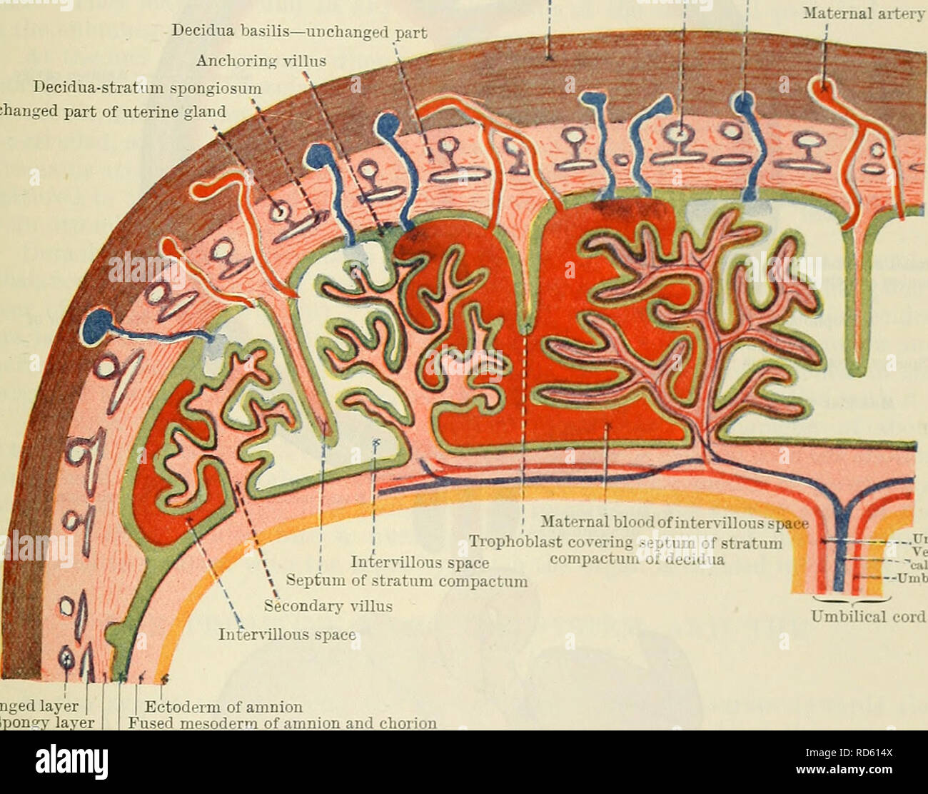
. Cunningham's Text-book of anatomy. Anatomy. THE PLACENTA. 61 of the uterus, until it is forced against the surrounding wall of the uterine cavity, where it fuses with the decidua vera, and thus the cavity of the uterus is obliterated. This fusion takes place towards the end of the second month, and as soon as it has occurred the discoid mass of placental tissue is continuous at its margin with the fused amnion, chorion, and decidua vera (Fig. 78). After the second month the fcetus lies in the amnion cavity, which is bounded by the fused chorion and uterine wall, except at the lower end of the uterus, where, over the onficium internum, the cavity of the body of the uterus communicates with the cavity of the neck of the uterus; there the amniotic cavity is bounded by a mem- brane formed by the fused amnion chorion lseve and the decidua capsularis only. And at the end of pregnancy this portion of the membrane is ruptured by the increased pressure of the amnion fluid produced by the contraction of the muscular wall of the uterus (Fig. 88). Unchanged part of uterine gland Muscular wall of uterus Maternal vein Decidua basilis—unchanged part Anchoring villus Decidua-stratum spongiosum Unchanged part of uterine gland Maternal artery. Unchanged layer | Spongy layer Ectoderm of amnion Fused mesoderm of amnion and chorion Compact layer Trophoblast of chorion Fig. 79.—Schema of Structure of Completed Pi.acf.nta. Completion of the Placenta.—It has already been stated that each secondary villus consists of a vascular mesodermal core covered by a cellular and a plasmodial layer of trophoblast, the latter lying next the maternal blood in the intervillous spaces. As development proceeds and the intervillous spaces become larger, the villi become longer and more complicated, and at the same time the cellular layer of the trophoblast largely disappears, until in the majority of the villi the plasmodial layer alone covers the vascular mesodermal core. In still later stages, deg