. Le coeur et l'aorte; études en radiologie clinique . ig. 53. BIGHT POSTEEIOE POSITION OBLIQUE, 50 DEGBEES.MODEEATE DILATATION DE L'AUEICLE GAUCHE INSUFFISANCE MITRALE l'histoire clinique de l'insuffisance mitrale presentsproblèmes, dont la solution de deux, au moins, est noteasy. La première est de savoir si le murmure systolique entendu atl'apex appartient simplement à la catégorie des anorganismurmures (Potain); la seconde est de reconnaître la cause de l'ofit, car le murmure peut être symptomatique d'une valvularlésion ou d'une insuffisance fonctionnelle. À cet égard, les indications fournies par semeiologie sont souvent
1669 x 1498 px | 28,3 x 25,4 cm | 11,1 x 10 inches | 150dpi
Informations supplémentaires:
Cette image peut avoir des imperfections car il s’agit d’une image historique ou de reportage.
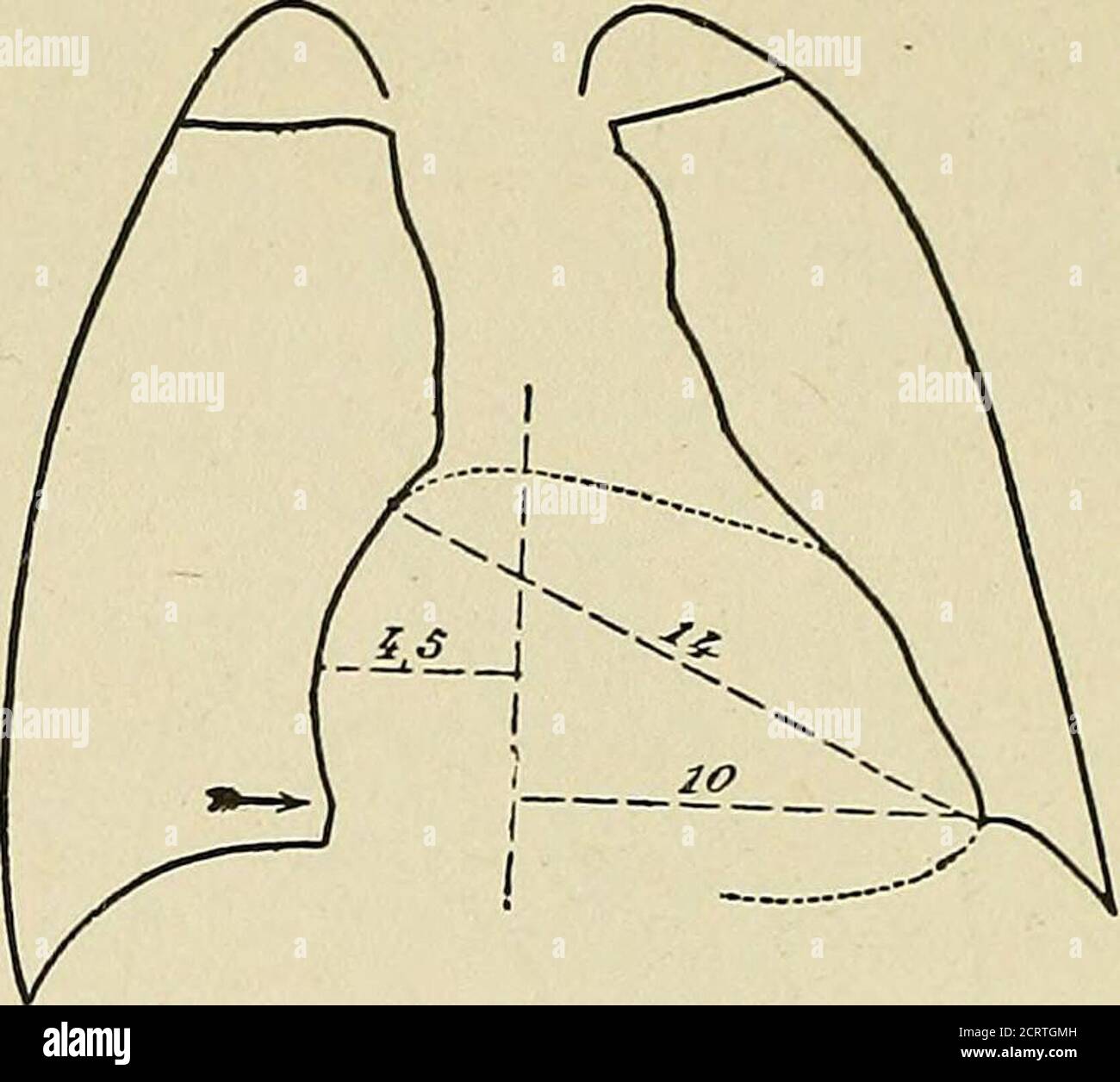
. The heart and the aorta; studies in clinical radiology . ig. 53. BIGHT POSTEEIOE OBLIQUE POSITION, 50 DEGBEES.MODEEATE DILATATION OF THE LEFT AUEICLE MITRAL INSUFFICIENCY The clinical history of mitral insufficiency presentsproblems, the solution of two of which, at least, is noteasy. The first is whether the systolic murmur heard atthe apex belongs simply to the category of anorganicmurmurs (Potain); the second is to recognize the cause ofit, as the murmur may be symptomatic of a valvularlesion or of functional insufficiency. In this respect theindications furnished by semeiology are often uncertainand radiological examination may be of very great assist-ance. The radiograms of a typical case of mitral insufficiencyof rheumatic origin which has not given rise to markedcirculatory disturbances will be studied first. This caseis a patient twenty-five years of age, with a loud murmurheard at the apex of the heart during the whole systole 88 THE HEART AND THE AORTA and transmitted toward the axilla. The affection beganin adolescence (Fig. 54).. Fig. 54. MITEAL INSUFFICIENCY WITH COMPENSATION.25 YEARS OF AGE MAN The form of the area of projection somewhat resemblesthat of a normal horizontal heart, resting on the dia-phragm. Its development, however, is clearly exagger-ated on the right. Moreover, on the screen, pulsationscould be seen in the vicinity of the diaphragmatic shadow(at the level of the arrow), which could only be causedby the right ventricle. The contour of the left ventricle appears normal, itsleft point not elevated. The apex lies at the level of theleft diaphragmatic shadow; it is not lowered, but pushedoutward and rather pointed. The changes seem to affect only the right heart areaand this is confirmed by the measurement of the diame-ters : longitudinal diameter, 14 cm.; horizontal diameter, 14.5 cm. The longitudinal diameter does not exceed the normalfor a man twenty-five years of age, of average weight, butthe horizontal diameter is 5 mm. greater tha