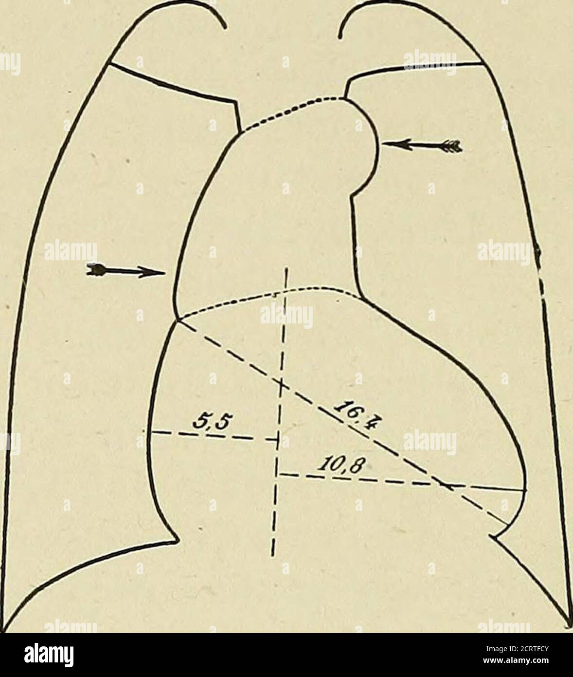
. Le coeur et l'aorte; études en radiologie clinique . ratages. Orthodiagramme 77 est d'un patient de quarante ans avec une sténose aortique grave, sans signe d'insuffisance cardiaque. Le point à noter ici est le dével-opment excessif du volume du coeur; le diam-mètre longitudinal mesure 17.5 cm.; l'horizontale, 17.8 cm.; les aortachows pas de lésion. Dans ce cas, le diagnostic clinique était apparent et la radioscopie confirmatoire. Figure 77. STÉNOSE AORTIQUE SANS AORTITIS. HOMME 40 ANS 112 LE COEUR ET L'AORTE ce n'est pas toujours le cas. Souvent, il est difficile de savoir whetheror
1505 x 1660 px | 25,5 x 28,1 cm | 10 x 11,1 inches | 150dpi
Informations supplémentaires:
Cette image peut avoir des imperfections car il s’agit d’une image historique ou de reportage.
. The heart and the aorta; studies in clinical radiology . racings. Orthodiagram 77 is of a patient forty years of age withserious aortic stenosis, with no sign of cardiac insuffi-ciency. The point to be noted here is the excessive devel-opment of the volume of the heart; the longitudinal diam-eter measures 17.5 cm.; the horizontal, 17.8 cm.; the aortashows no lesion. In this case the clinical diagnosis wasapparent and radioscopy confirmatory. Fig. 77. AORTIC STENOSIS WITHOUT AORTITIS. MAN 40 YEARS OF AGE 112 THE HEART AND THE AORTA It is not always so. Often it is difficult to know whetheror not aortic stenosis exists, for the systolic murmur atthe base is difficult to interpret, and hypertrophy of theleft ventricle always present in this disease is not suffi-ciently marked to be obtained by palpation or percussion.It is in such cases that radiology is most effective, andseveral times in debatable cases, aortic stenosis had tobe determined by the single fact that screen examinationdemonstrated the presence of a left ventricular hyper-trophy.. Fig. 78. AORTIC STENOSIS. DILATATION AND FORCIBLE PULSA-TION OF THE AORTA. YOUTH 17 YEARS OF AGE This examination leads to other findings which have animportant bearing on the prognosis of aortic stenosis, which varies according as the lesion is simple or accom-panied by more or less extensive changes of the aorta.These findings should be interpreted with great care, asthe following case shows. A youth seventeen years of age affected with aorticstenosis as shown by a systolic murmur at the base. Theorthodiagraphic tracing (Fig. 78) confirmed the diagno-sis, for all the objective signs were characteristic. On VALVULAR AFFECTIONS 113 superficial examination, it might have been thought thatthere were at the same time marked lesions of the vessel, which would have given an unfavorable prognosis. In thefrontal position, an evident enlargement of the arch wasobserved, its total transverse diameter being 7.5 cm.instead of 4 or 5 cm., t